The research results were published in Forensic Science International: Genetics.
Forensic DNA is known as the king evidence in courtroom trial. However, with the increasing ability of criminal suspects to evade investigation, the DNA evidence left at the crime scene is very limited and contains a large amount of inhibitor substances. Therefore, how to extract enough DNA from trace evidence samples for genetic marker typing has always been a challenge in forensic DNA analysis and testing.
In this study, researchers developed novel Chitosan@Beads and a modified lysis buffer.
"It can adsorb negatively charged DNA through electrostatic interactions," said ZHU Cancan, member of the team.
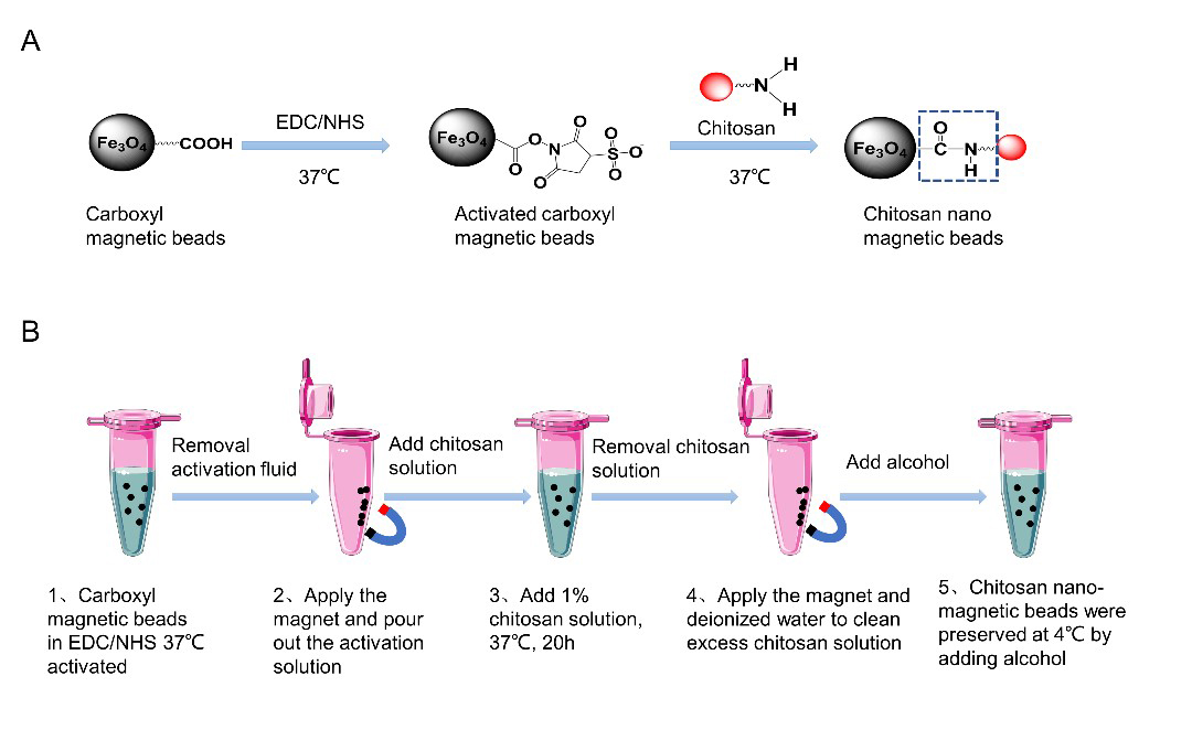
They used 1% (w/v) of chitosan (75% deacetylation degree) to modify the 50 nm magnetic nanoparticles and gained the Chitosan@Beads, which theoretically carried positively charged in the pH = 5 of lysis buffer.
To simulate real crime sense, a set of simulated samples, containing 20 mg/μL of humic acid, pigments, iron ions (Fe2+, Fe3+), and the coexistence of the above three substances, were prepared. Human bronchial epithelial cells were mixed with the 200μL of the above simulated samples for DNA extraction. 400μL of lysis buffer, 20 μL of proteinase K (10 mg/mL) and 20μL of Chitosan@Beads solution (20 mg/mL) was used for cell disruption and DNA enrichment. The results showed that the sensitivity of DNA extraction using Chitosan@Beads was superior to commercial reagent kits, with a detection limit of 10 cells.
Furthermore, for complex trace samples from case materials, the STR loci rate of the Chitosan@Beads was 97.9%, exhibiting a higher success rate in STR (short tandem repeat) analysis, an important technique used in forensic investigations.
This research has broad prospects for application in the fields of forensic DNA identification, ancestral analysis, martyr remains identification and identification of victims of major disasters.

Preparation of Chitosan@Beads. A: schematic diagram of chitosan-modified magnetic nanobeads; B: Schematic diagram of Chitosan@Beads modification steps. (Image by ZHU Cancan)

